Welcome to the Center for Radiological Research

Our Mission
The mission of the Center for Radiological Research (CRR) is to be on the forefront of radiological science and its applications in clinical medicine, public health, and national defense.
First International Congress on Far-UVC Science and Technology (ICFUST)

June 14-16 at Columbia University in New York City.


News
- February 27, 2024
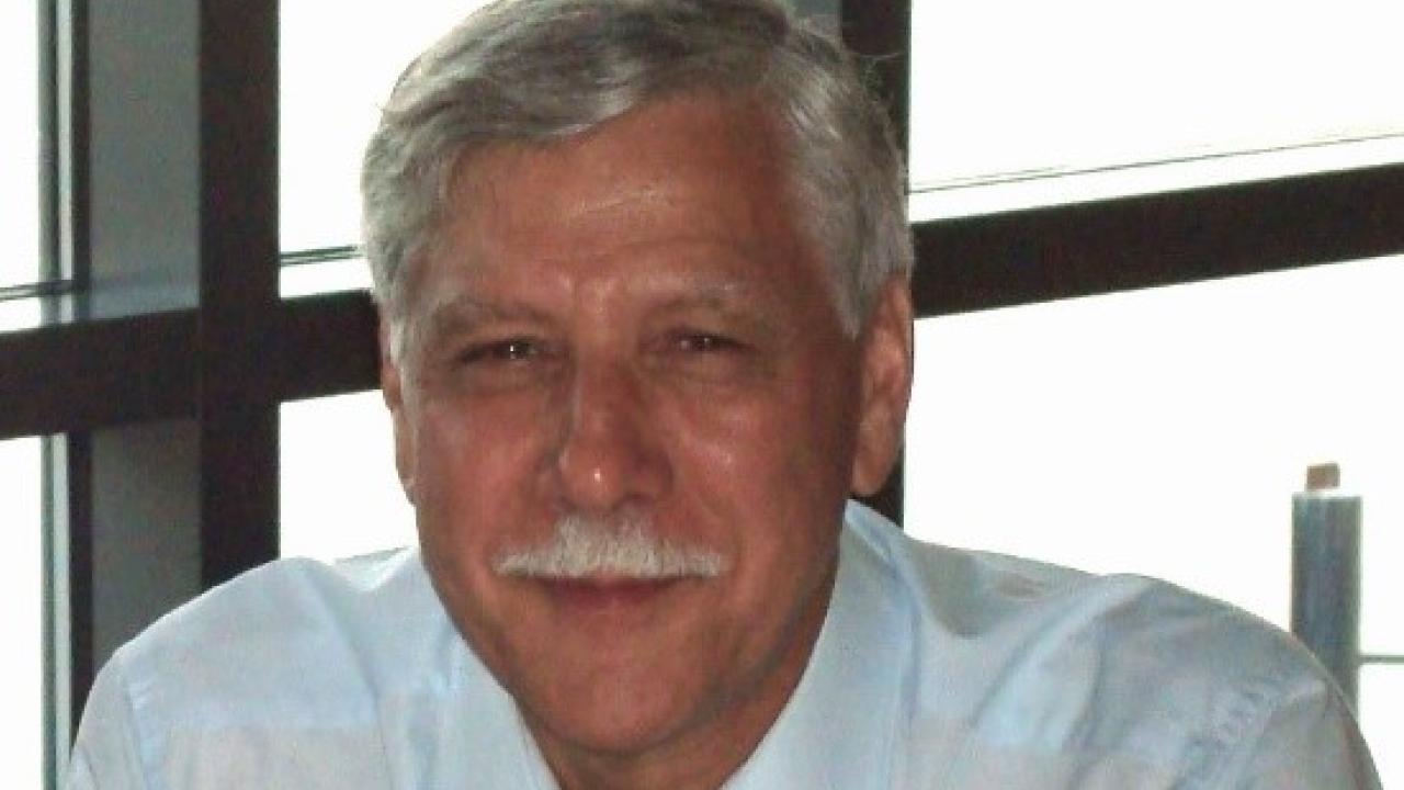
Steve Marino a long time member of the CRR and RARAF, suddenly passed on January 4th, 2024. This is a memorial tribute to Steve.
- June 29, 2023
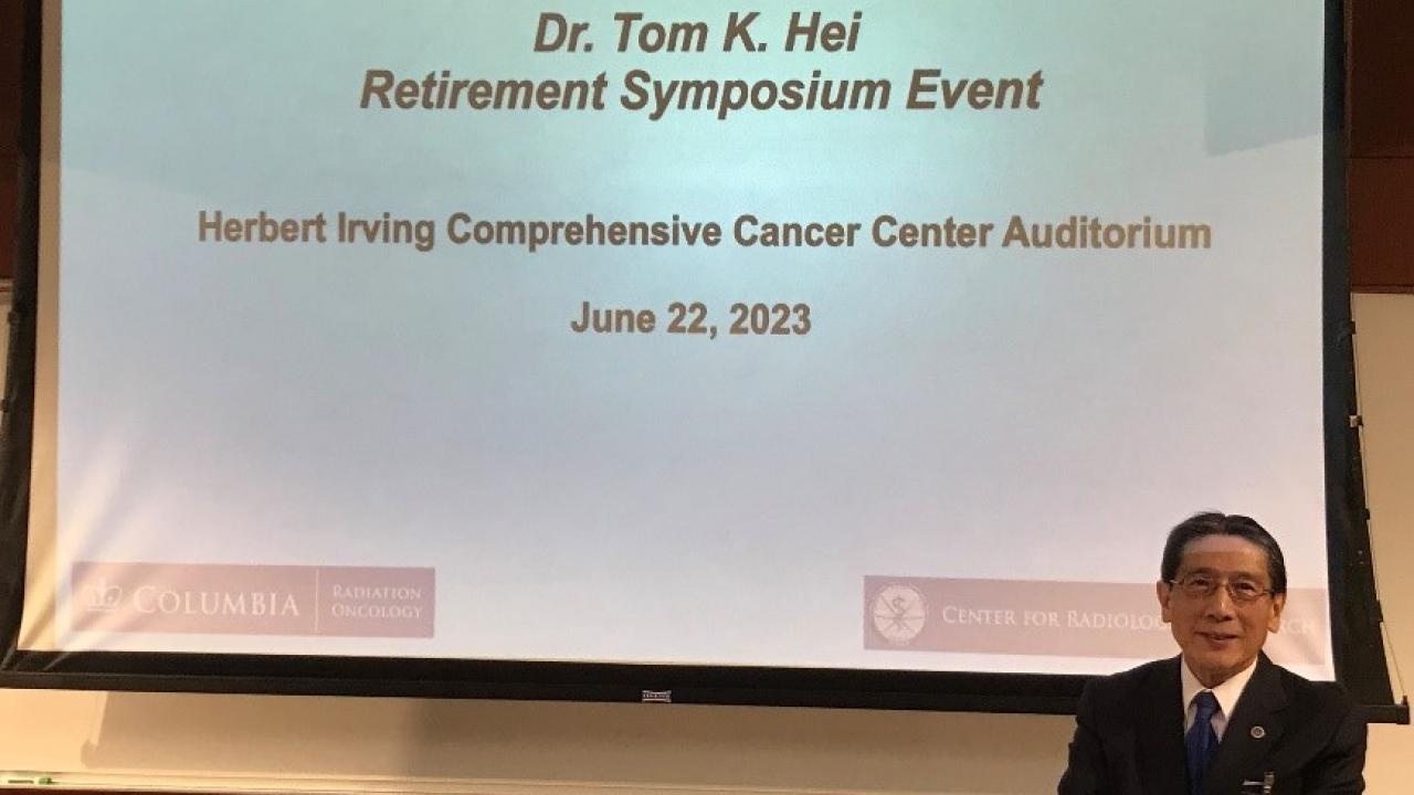
Dr. Tom Hei retired from Columbia University Irving Medical Center July 1st, 2023
Topic
Cancer - March 24, 2023
The Center for Radiological Research at Columbia University will be hosting the First International Congress on Far-UVC Science and Technology on June 14th-16th, 2023.
Topic
Public Health
Events
There are currently no upcoming events.
Support Our Mission

Donate to the CRR Training and Education Fund to help train a new generation of talented and committed young radiation scientists.
Radiation Oncology

The Center for Radiological Research is a part of the Columbia University Irving Medical Center, Department of Radiation Oncology